


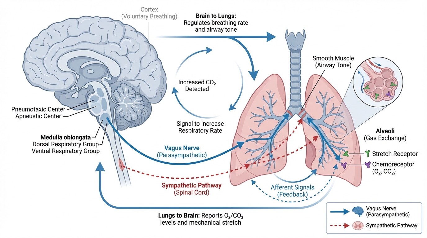
To master neuro-ventilation, you first need to appreciate one of the most fundamental relationships in critical care: the connection between the lungs and the brain. Think of it like a finely tuned hydraulic system. The brain, encased in the rigid skull, is incredibly sensitive to pressure changes. The lungs, managed by the ventilator, are a powerful source of that pressure. Every breath delivered by the ventilator can alter blood flow and pressure within the head.
Understanding this delicate cause-and-effect relationship is the foundation of safe and effective care. It’s not enough to just manage oxygen and carbon dioxide levels; you have to see the bigger picture. You must anticipate how each ventilator adjustment—from a change in tidal volume to an increase in pressure—will ripple through the body and impact the injured brain. Let’s dive into the core principles that govern this critical connection.
The Skull: A Closed Box with Strict Rules
The starting point for understanding brain-lung interactions is a concept from the 1780s that is still central to neurocritical care today: the Monro-Kellie doctrine. This principle states that the skull is a rigid, fixed-volume container. Inside this container are three main components:
- Brain Tissue (about 80% of the volume)
- Blood (about 10%)
- Cerebrospinal Fluid (CSF) (about 10%)
Because the skull cannot expand, if the volume of one component increases, the volume of one or both of the other components must decrease to maintain a stable intracranial pressure (ICP). For example, when a brain injury causes swelling (edema), the body first tries to compensate by pushing out some CSF and venous blood.
However, this compensation has its limits. Imagine a cup filled to the brim with water. You can add a few more drops, and the water will bulge at the surface before spilling. But once you reach that limit, even a single extra drop causes an overflow. The injured brain is on a similar “pressure-volume curve.” Once its compensatory mechanisms are exhausted, even a tiny increase in volume—like a small amount of extra blood flow—can cause a dramatic, dangerous spike in ICP. This is where the ventilator comes in, as it directly influences the volume of blood inside the skull.
Chemical Control: How Gases Steer Blood Flow
The most powerful tool the ventilator has to influence the brain is its ability to control blood gas levels, specifically carbon dioxide (PaCO₂) and oxygen (PaO₂). These gases are potent regulators of the blood vessels in the brain.
Carbon Dioxide: The Brain’s Accelerator and Brake
Carbon dioxide is the most influential chemical modulator of cerebral blood flow. It diffuses easily across the blood-brain barrier, changing the local pH and causing blood vessels to either expand or contract.
- Hypercapnia (High PaCO₂): When CO₂ levels rise, it causes cerebral vasodilation—the blood vessels in the brain widen. This increases the total volume of blood within the skull. In a healthy person, this isn’t a problem. But in a patient with a swollen brain who is already on the edge of their pressure-volume curve, this extra blood volume can be the “single drop” that causes a catastrophic rise in ICP.
- Hypocapnia (Low PaCO₂): Conversely, when CO₂ levels fall, it causes cerebral vasoconstriction—the blood vessels narrow. This reduces blood volume in the skull and rapidly lowers ICP. For decades, this was the go-to strategy for managing brain injuries. However, we now know it comes at a steep price. This constriction can become so severe that it chokes off blood flow, leading to cerebral ischemia, or a “stroke” within the injured tissue.
Think of PaCO₂ as the accelerator and brake for cerebral blood flow. Your goal is to keep the foot steady, maintaining a normal PaCO₂ level (typically 35-45 mmHg). Stepping too hard on the gas (hypercapnia) or slamming on the brakes (hypocapnia) can both lead to disaster.
Oxygen: A Lifeline with Limits
The brain is highly dependent on a constant supply of oxygen. Its response to oxygen levels is primarily a survival mechanism.
- Hypoxia (Low PaO₂): When oxygen levels drop below a critical threshold (around 60 mmHg), the brain triggers potent vasodilation as a desperate attempt to increase oxygen delivery. This dramatically increases blood volume and ICP. Avoiding hypoxia is a non-negotiable rule in neurocritical care.
- Hyperoxia (High PaO₂): For a long time, it was thought that more oxygen was always better. We now know that’s not true. Excessive oxygen can cause a mild vasoconstriction, reducing cerebral blood flow. More importantly, in the context of reperfusion after an injury (like a stroke), very high oxygen levels can create harmful molecules called reactive oxygen species that worsen neuronal damage. The goal isn’t super-saturation; it’s a safe and steady state of normoxia.
Mechanical Pressure: The Ventilator’s Squeeze
Beyond chemistry, the ventilator exerts a direct mechanical force that affects the brain. The positive pressure it generates in the chest to inflate the lungs doesn’t just stay in the chest. It gets transmitted to the entire circulatory system, impacting how blood gets back to the heart from the head.
This is known as the “Starling resistor” effect. The veins that drain blood from the brain are soft and have no valves. They pass through the chest on their way back to the heart. When you apply positive pressure in the chest with a ventilator (especially with Positive End-Expiratory Pressure, or PEEP), you increase the intrathoracic pressure. This can squeeze those veins.
If the pressure in the chest (Central Venous Pressure, or CVP) becomes higher than the pressure in the head (ICP), it acts like a dam, obstructing the outflow of blood from the brain. This creates a backlog, causing venous congestion in the head and increasing intracranial pressure.
A crucial nuance here is lung compliance. If a patient has stiff, non-compliant lungs (like in ARDS), the lungs act as a shield, absorbing much of the ventilator pressure and preventing it from being fully transmitted to the chest cavity. In these cases, higher levels of PEEP may have less impact on ICP. Conversely, in a patient with healthy, compliant lungs, that pressure is transmitted much more easily, posing a greater risk to the brain.
Finally, we must always consider Cerebral Perfusion Pressure (CPP), which is the net pressure that drives blood flow to the brain. The formula is simple but vital:
CPP = Mean Arterial Pressure (MAP) – Intracranial Pressure (ICP)
Mechanical ventilation can harm CPP from both ends. As we’ve seen, it can increase ICP. It can also decrease MAP by reducing the amount of blood returning to the heart, thus lowering cardiac output. A drop in MAP directly lowers CPP, starving the brain of the blood it needs. Every ventilator change requires you to ask: how will this affect both my ICP and my MAP?
By understanding these fundamental brain-lung interactions, you move from simply operating a ventilator to becoming a true neuro-pulmonary physiologist. You can begin to anticipate the downstream effects of your actions, making informed decisions that protect both the lungs and the brain.


1 thought on “Brain-Lung Interactions: The Critical Connection”
This is great information. Keep it coming please!!