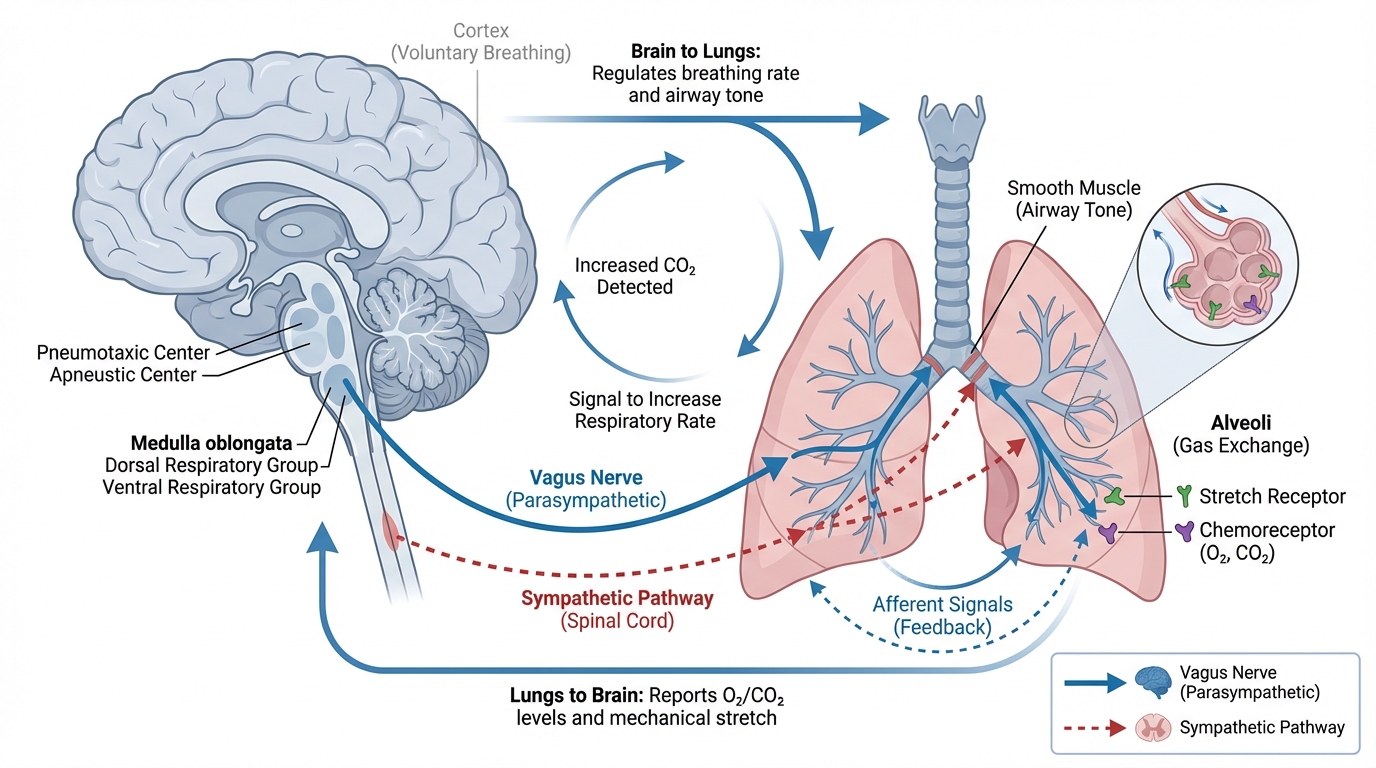


How exactly do the lungs and brain talk to each other during critical care? It might seem like they operate in different worlds—one managing air, the other thoughts. But in a patient with a brain injury, these two organs are in constant, critical communication. The ventilator isn’t just a breathing machine; it’s a device that sends powerful messages to the brain with every single breath.
Understanding this physiological dialogue is like learning the secret language of the ICU. It allows you to see how a simple adjustment on the ventilator can create ripples that affect the pressure inside the skull. This knowledge is your foundation for moving beyond just following protocols to truly understanding why you’re making each decision. So, let’s tune into that conversation and decode the three main ways the lungs and brain interact: through pressure, chemistry, and flow.
The Skull: A Closed Box with Strict Rules
To get started, we need to travel back to the 1780s and revisit a core principle of neurocritical care: the Monro-Kellie doctrine. Think of the skull as a sealed, rigid box. It’s like a high-security vault that cannot expand. Inside this vault, there are three main things:
- Brain Tissue (about 80% of the space)
- Blood (about 10%)
- Cerebrospinal Fluid (CSF) (about 10%)
Because the total volume inside this box is fixed, if one component swells, another must shrink to make room and keep the pressure stable. When a brain injury occurs, the tissue swells with edema. The body’s first response is to compensate by pushing out some CSF and venous blood. It’s the body’s natural pressure-relief system.
But this system has its limits. Imagine a water balloon filled almost to its breaking point. You can poke it gently, and the water will shift around. But if you add even a little more water, the balloon will burst. The injured brain exists on a similar “pressure-volume curve.” Once its ability to compensate is maxed out, it’s at that breaking point. Even a tiny increase in volume—like a small surge of blood—can cause intracranial pressure (ICP) to spike dangerously. This is where the ventilator becomes a major player, as it directly influences the amount of blood inside that closed box.
Chemical Control: How Gases Steer Blood Flow
The ventilator’s most direct line of communication with the brain is through its control of blood gases. Specifically, the levels of carbon dioxide (PaCO₂) and oxygen (PaO₂) in the arterial blood are powerful signals that tell the brain’s blood vessels what to do.
Carbon Dioxide: The Brain’s Accelerator and Brake
Carbon dioxide is the ultimate influencer of cerebral blood flow. It slips across the blood-brain barrier with ease, changing the pH of the local environment and causing blood vessels to either open up or clamp down.
- Hypercapnia (High PaCO₂): When CO₂ levels in the blood rise, it signals the cerebral blood vessels to dilate, or widen. This is called vasodilation. In a healthy person, this is no big deal. But for your patient with brain swelling, it’s a huge problem. That vasodilation brings more blood into the skull, increasing the volume inside the fixed box. If the brain is already on that steep part of the pressure-volume curve, this extra blood volume can trigger a catastrophic rise in ICP.
- Hypocapnia (Low PaCO₂): On the flip side, when you “blow off” CO₂ and its levels drop, it causes cerebral vasoconstriction. The blood vessels narrow, reducing the amount of blood in the skull and quickly lowering ICP. For decades, this was the standard treatment for high ICP. The problem, as we now know, is that this constriction can be too effective. It can choke off blood flow to the point of causing cerebral ischemia—essentially, a stroke within the already injured brain tissue.
Think of PaCO₂ as the gas pedal for cerebral blood flow. You want to keep a steady foot, maintaining a normal level (typically 35-45 mmHg). Slamming on the gas (hypercapnia) or the brakes (hypocapnia) can both lead to disaster. Your job is to find that perfect cruising speed.
Oxygen: A Lifeline with Limits
The brain is an oxygen hog, and its response to changing O₂ levels is all about survival.
- Hypoxia (Low PaO₂): When the arterial oxygen level drops below a critical point (around 60 mmHg), the brain sounds the alarm. It triggers widespread vasodilation as a last-ditch effort to pull in more oxygen-rich blood. This floods the intracranial space with blood, causing ICP to skyrocket. Avoiding hypoxia is one of the absolute, non-negotiable commandments of neurocritical care.
- Hyperoxia (High PaO₂): For a long time, the thinking was, “if some oxygen is good, more must be better.” We now know this isn’t the case. While not as dramatic as hypoxia, flooding the body with excess oxygen can cause mild vasoconstriction in the brain, slightly reducing blood flow. More importantly, in situations like recovery from a stroke, too much oxygen can create harmful molecules called reactive oxygen species, which can add insult to injury by damaging neurons. The goal isn’t to blast the brain with oxygen; it’s to ensure a safe and steady supply, a state known as normoxia.
Mechanical Pressure: The Ventilator’s Squeeze
The final piece of the puzzle is the direct mechanical force of the ventilator. The positive pressure used to inflate the lungs doesn’t just stay in the chest; it sends pressure waves throughout the entire circulatory system. This directly affects how blood gets from the head back to the heart.
This phenomenon is often described using the “Starling resistor” model. The veins that drain blood from the brain are soft, collapsible tubes without valves. To get back to the heart, they must pass through the chest. When you use a ventilator to apply positive pressure to the lungs—especially with Positive End-Expiratory Pressure (PEEP)—you increase the pressure inside the chest (intrathoracic pressure).
This increased pressure can squeeze the draining veins. If the pressure in the chest (measured as Central Venous Pressure, or CVP) rises above the pressure in the head (ICP), it’s like putting a clamp on a garden hose. It obstructs the outflow of blood from the brain. Blood backs up, creating venous congestion inside the skull and raising ICP.
There’s an important variable here: lung compliance. If your patient has very stiff, sick lungs (as in Acute Respiratory Distress Syndrome, or ARDS), the lungs act like a sponge, absorbing much of the ventilator pressure. This shields the chest cavity, and less pressure is transmitted to the veins. In this case, higher levels of PEEP might be well-tolerated. But if your patient has healthy, compliant lungs, that pressure is transmitted much more easily, posing a greater risk of impeding drainage from the brain.
Finally, all these factors come together to influence Cerebral Perfusion Pressure (CPP). This is the net pressure that actually pushes blood and oxygen into the brain tissue. The formula is simple but crucial:
CPP = Mean Arterial Pressure (MAP) – Intracranial Pressure (ICP)
Mechanical ventilation can attack this equation from both sides. As we’ve seen, it can raise ICP. It can also lower MAP by squeezing the heart and reducing the amount of blood returning to it, thereby lowering cardiac output. A drop in MAP directly translates to a drop in CPP, potentially starving the brain. When you change a ventilator setting, you have to ask yourself: how will this affect both sides of the CPP equation?
By understanding these core brain-lung interactions, you elevate your practice. You are no longer just a technician setting dials; you are a physiologist managing a complex, dynamic system. You can anticipate the consequences of your actions, making thoughtful choices that protect both the lungs and the brain, and guiding your patients toward the best possible outcome.